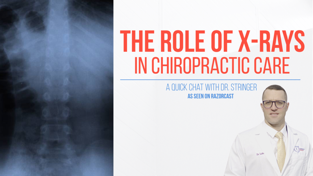
We use X-rays because to see is to know. And if we’re going to work with the spine and the nervous system, I want to see exactly what’s going on within that spine. For us, 95% of the time we are going to take an X-ray because we want to see exactly what that spine looks like. I want to know what the spine looks like to obviously develop a plan that’s going to work best for that spine, if that includes adjustments, traction, physical therapy, soft tissue, or all in between. It’s like building a house. You don’t build a house without blueprints. Otherwise, you’re guessing. Same with working with the spine.
Taking X-rays is also going to make sure that we’re being super diligent in evaluating the health of the spine on a neuro and a physiological level. And also, how are we going to adjust that spine? If we have a reduction in the curve or any shifts, we want to know about it because how we prescribe treatment involves spinal traction. We can objectively change the shape of that patient’s spine, which, research supplements, improves objective outcomes long-term. Also, there might be a growth or a tumor or an infection within the spine the patient doesn’t know about it, and we can catch on X-rays.
We take X-rays, we evaluate them on our own, but we also refer them out to radiologists to get a second set of eyes on them. I do that to be super thorough; not all chiropractors do that. Some do, some don’t, but chiropractors within their scope of practicing license are able to take X-rays and read them comprehensively in the state of Illinois. It might vary in a couple of different states, but in our state, you can absolutely do that.
Chiropractors take a massive amount of radiology curriculum and board examinations ranging from how to take them to how to review them and then going through all the pathology within the spine. Actually, chiropractors can do a crossover where they’re called a DACBR where they are actually clinically trained like radiologists to read images, MRI, CTs, X-rays, et cetera.
But before we take an X-ray, we’re going to do a detailed consult and a really detailed examination. There’s going to be an orthopedic, neurological and functional exam. I’m going to ask you to go through range of motion. I’m going to perform an orthopedic test, watch those structures move, and see where you are weak and deficient. I’m going to perform a neurological exam and see if the nerves in the spine are doing their job. I’m going to functionally evaluate the joints. That exam will then dictate if we need X-rays and then we take them ourselves.
There are many different kinds of X-rays you can take like specific films for specific conditions. But typically for us, it is going to be the standard front to back, side and then movement film if we see any big kind of trauma or any pathology on there.
A typical series of X-rays for your lower back is going to be what we call A to B from the front to the back. You’re going to get all the pelvic, the sacral and low back anatomy in there. And then what we call a lateral film where we take it from the side.
Now, if you have had a big trauma, been involved in a car accident, we might take what we call a flexion film and an extension film, where we bend to one way and we bend to the other, or we bend forwards, or we bend backwards. We do that to look for ligament damage because joints will shift and move on the X-ray.
When we’re looking at the X-rays, what are we looking for? We are looking for good joint alignment, good spine alignment, good bone health, good disc health. Have we got occlusion within the nerve canal? Do we have any pathology in there or any infection or do we have any soft tissue damage? Do we have rotation in the knee? For the shoulder, same thing when we have that forward head posture, do the shoulders round in, does that shoulder blade wing out? Do we have impingement in the shoulders? Do we have degeneration?
For example, I had a patient who was younger, active, and his pain level was really low and had not been going on for a massive amount of time. We shot some X-rays and his spine was massively shifted, an inch to the left, inch forward, and reverse in his curve. His main concern was headaches and the atlas bone, which is the top bone in your neck, was actually poking up into his skull. I need to know that. If I’m going to adjust that bone, I want to make sure that I’m adjusting it appropriately.
So, X-rays play a role in putting together the puzzle of figuring out what’s going on for that patient and then the best type of treatment for them. It’s a great, great tool. And it’s an objective tool to figure out the best course of treatment for that patient.
